This model was designed and tested as part of the 3D4VIP project.
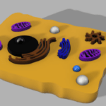
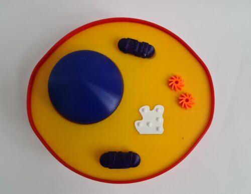
Animal cell with different organelles:
-cell membrane
-cytoplasm
-nucleus
-mitochondria
-free ribosomes
-centrioles

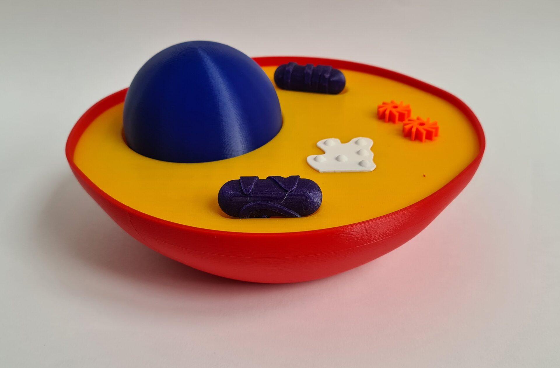
For printing the animal cell I would suggest to use different colours for different parts of the model. Depending on the quality of the printer you can use 0.10, 0.15 or 0.2 mm layer height for the different parts. For the smaller parts I would definitely use 0.1 mm layer height.
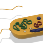
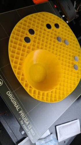
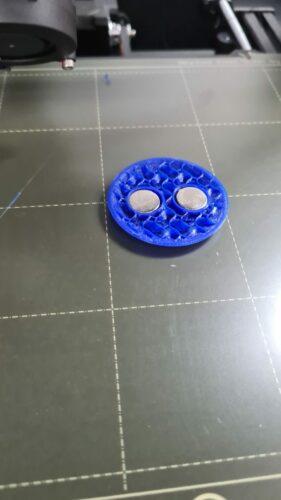
Also, the model uses magnets to keep the parts in place. For using magnets in the lower cytoplasm part you need two moments were you pause the print to glue the magnets into place. First, the magnet holes are created for the nucleus:
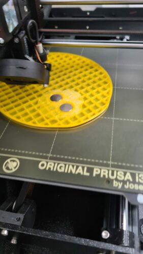
The second pause for the printer is when the magnet holes for the other organelles have been finished.
You can also add the magnets in the nucleus file by creating a pause after the two holes are created:
I printed all the organelles with metal-filled filament so they are attracted by the magnets. This means they all have the same color, but it helps a lot with keeping all the parts of the model together.